Acoustic neuroma is the most common type of brain tumor. It is noncancerous and grows on a tiny nerve that is located near facial nerves between the inner ear and brainstem.
An acoustic neuroma occurs on the eighth cranial nerve. It consists of three nerves that link the eardrum to the brain, including the cochlear nerve (carries hearing information) and left and right nerves (carry balance signals from the inner ear to the brain). Schwann cells (neurilemma cells) protect these nerves. However, an acoustic neuroma causes a tumor to grow on Schwann cells. And if the tumor persists, it will compress the brainstem. Thus, an acoustic neuroma can be life-threatening.
Acoustic neuromas can develop at any time. Yet, those who understand acoustic neuromas can keep an eye out for the symptoms associated with these tumors. From here, they can identify these symptoms in their early stages. And if necessary, these individuals can seek out medical treatment to address their acoustic neuroma symptoms before they cause long-lasting health problems.
What is an Acoustic Neuroma?

Acoustic neuromas are vestibular schwannomas, i.e. noncancerous tumors that form in the ears. They look similar to tumors that can affect the middle and inner ear portions. Acoustic neuromas can cause symptoms comparable to these tumors as well.
An acoustic neuroma generally grows slowly in the area where the central nervous system transitions into the peripheral nervous system. In many instances, this type of tumor stops growing altogether.
In rare instances, acoustic neuromas continue growing large over an extended period of time. As these instances occur, the tumors won’t necessarily damage tissue. Rather, the tumors compress the nerves that promote facial sensation and facial muscle movement.
If acoustic neuromas go undiagnosed and untreated, they can cause severe compression of the eight cranial nerve that impacts hearing and balance. Thus, individuals dealing with acoustic neuromas may be prone to facial numbness. They are also susceptible to hearing and balance problems.
How Common Are Acoustic Neuromas?
Acoustic neuromas are rarely inherited from a parent. However, an acoustic neuroma caused by neurofibromatosis type II (NF 2) is more common in young patients and those with a family history of neural tumors.
The National Institutes of Health (NIH) has performed a variety of acoustic neuroma and acoustic neuroma facial paralysis studies, and some of their study findings include:
- Acoustic neuromas comprise roughly 6% of all intracranial tumors, 30% of brainstem tumors, and 85% of tumors in the cerebellopontine angle (CPA) region of the brain.
- 10% of acoustic neuromas are meningiomas (noncancerous tumors that surround the brain and spinal cord).
- Only about one out of every 100,000 acoustic neuromas are diagnosed annually in the United States.
- Each year, there are roughly 2,000 to 3,000 new acoustic neuroma cases reported in the United States.
- Among patients who suffer from hearing asymmetry, only about one in 1,000 is dealing with an acoustic neuroma.
The treatment approach to facial paralysis in this patient population depends on the intraoperative surgical findings. A conservative approach is prudent if the surgeon who performs an acoustic neuroma feels that the nerve was saved during the operation.
Acoustic neuroma patients will often require eye care and temporary measures such as suture suspensions to prevent unwanted complications and improve oral issues. The risk of facial paralysis with acoustic neuroma surgery is between 4-15%. Tumor size, surgeon experience, and approach for surgery are important factors to determine the success of an acoustic neuroma procedure.
Acoustic Neuroma Causes
Most individuals diagnosed with an acoustic neuroma have no apparent risk factors. One confirmed environmental cause of the condition is exposure to high dose radiation, especially in the face or neck. Contrary to popular belief, cell phone use is not a contributor to the development of an acoustic neuroma. Acoustic neuromas may also be caused by continuous exposure to loud music or work-related noise.
A special group of individuals face a high risk of developing acoustic neuromas. These are individuals with a rare genetic disorder called neurofibromatosis type 2 (NF2), which accounts for 5% of all cases of acoustic neuromas. NF2 is caused by a malfunctioning tumor suppressor gene on chromosome 22, leading to the growth of benign (noncancerous) tumors on the nerves that control balance in the inner ear. Although these tumors are benign, they can cause serious hearing and balance complications. NF2 can be familial and having a parent with the condition also increases your chances of developing an acoustic neuroma, because it is a dominant gene. In other words, you have a 50-50 chance of developing NF2 if one of your parents has been diagnosed with the condition.
Acoustic Neuroma Symptoms
More than 95 percent of acoustic neuroma patients suffer hearing loss. In addition, roughly 90 percent of acoustic neuroma patients encounter a gradual, one-side hearing impairment. Among patients who suffer a hearing impairment due to acoustic neuroma, nearly two-thirds deal with a high-frequency sensorineural pattern. Meanwhile, the remainder frequently suffer from low-frequency hearing loss, i.e. a form of Meniere’s disease.
In some instances, acoustic neuroma patients deal with “cookie bite” pattern, an indication of congenital hearing loss. Sudden hearing loss also occurs among 25 percent of acoustic neuroma patients but is rarely attributed directly to acoustic neuroma. Conversely, sudden hearing loss is attributed to an acoustic neuroma patient only about 1 percent to 5 percent of the time, as there are many causes of sudden hearing loss. Furthermore, hearing remains normal in about 11 percent of acoustic neuroma patients. Tinnitus (perception of noise ringing in the ears) also may be attributed to acoustic neuroma; it often is unilateral and confined to the affected ear.
Due in part to the origin of acoustics in the vestibular nerve, about 20 percent of acoustic neuroma patients will experience vertigo. In many cases, vertigo is more common in patients with smaller tumors than larger ones. Meanwhile, a lack of balance affects approximately 70 percent of acoustic neuroma patients who experience vertigo. Most of these patients will have large tumors. Loss of use of one or more of the arms and legs may occur but are unusual.
Acoustic neuroma facial paralysis may occur if a large tumor is present. In some situations, carbamazepine medication for neuralgia may enable a patient to limit acoustic neuroma facial nerve damage. An acoustic neuroma patient also may experience headaches if he or she is dealing with a large tumor.
If the nerve was cut intraoperatively or if the facial palsy does not resolve, then a more active approach must be taken. Definitive eyelid reconstruction with a gold weight or palpebral spring must be considered. Facial reanimation as discussed in other sections of this website must then be utilized. The following section is a more detailed discussion about acoustic neuroma diagnosis.
Acoustic Neuroma Diagnosis
The diagnosis of acoustic neuroma usually involves a detailed physical examination to check your hearing and balance as well as the reflexes, strength, and sensation in your face and upper and lower extremities. Routine blood tests are generally not required to diagnose acoustic neuromas. In some cases, the auditory brainstem evoked response (ABR) is used as a screening test for acoustic neuroma. The definitive diagnostic imaging study for patients with suspected acoustic neuromas is gadolinium-enhanced magnetic resonance imaging (MRI) scanning. The procedure provides a highly detailed picture of the brain. The gadolinium, a type of contrast agent, is injected into a vein and is crucial in helping detect acoustic neuroma tumors as small as 1 to 2 mm in diameter. The scanning procedure is painless and takes about 30 minutes. Conversely, computerized tomography (CT) scanning with contrast can also be used to detect medium- to large-size acoustic neuroma tumors but fail to detect any tumors smaller than 1 to 1.5 cm in diameter. The study provides a three-dimensional (3D) picture of the brain and associated structures. It also is pain-free and takes almost 30 minutes to complete.
The procedures described will give your physician the best possible information regarding the type, position, and size of the acoustic neuroma tumor for the formulation of a highly personalized treatment plan.
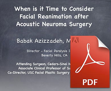
When is it time to consider facial reanimation after acoustic neuroma surgery?
View Dr. Azizzadeh’s presentation for the Acoustic Neuroma Association on facial paralysis treatment options following acoustic neuroma surgery. Dr. Azizzadeh discusses all of the current facial reanimation treatment options, including the management of partial paralysis and synkinesis.
Acoustic Neuroma Treatment Options
There are four distinct treatment options for an acoustic neuroma:
- Medical treatment or taking a “wait and see” approach (conservative management)
- Acoustic neuroma removal surgery
- Gamma-knife radiosurgery
- Cochlear implantation
Roughly 25 percent of acoustic neuromas are treated with medical management, which consists of:
- Periodic monitoring of the patient’s neurological status
- Use of hearing aids when appropriate
- Periodic imaging studies
Acoustic neuromas develop slowly, and there are no current medications that have been shown to limit acoustic neuroma growth. A patient may use serial audiometry and/or MRI scans to track an acoustic neuroma. After an acoustic neuroma is diagnosed, an MRI may be obtained at six months, followed by annual MRIs.
The threat of acoustic neuroma can decline based on an individual’s age. For example, an older acoustic neuroma patient may continue to take his or her current medications. In this instance, the acoustic neuroma may be unlikely to impact the patient’s expected lifespan.
Like any surgery, there are risks associated with an acoustic neuroma procedure. An MRI sometimes is unable to show how quickly an acoustic neuroma is growing, and the tumor could cause a patient to suffer hearing loss. If this happens, a patient may no longer be a viable candidate for a hearing preservation procedure.
A recent study indicated that roughly 10 percent to 43 percent of acoustic neuroma patients who were evaluated for about two years lost “useful” hearing. Typically, 75 percent of acoustic neuromas will display visible growth of approximately 1.5 mm over the course of one year. Keep in mind, however, that some acoustic neuromas will grow faster than others.
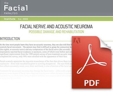
Possible Damage and Rehabilitation
For those diagnosed with an acoustic neuroma, there are many aspects of treatment. In this article, Dr. Azizzadeh discusses the nuances of treating an acoustic neuroma while preserving facial nerve function.
What Is Acoustic Neuroma Surgery?
Currently, about 50 percent of acoustic neuromas receive surgical treatment. Acoustic neuroma surgery often is a preferred choice for patients because it may prevent fatal complications associated with tumor growth and help an individual preserve his or hearing. The surgery usually is performed by a neurotologist (specialized otolaryngologist), neurosurgeon and other surgeons at an academic center.
There are several types of acoustic neuroma procedures:
- Retrosigmoid/Suboccipital: Serves as a posterior approach that involves acoustic neuroma removal through the skull.
- Translabyrinthine: Involves acoustic neuroma removal through the inner ear, commonly resulting in hearing loss.
- Middle Fossa: Offers acoustic neuroma removal through the skull, improving a patient’s chances of preserving his or her hearing.
Each acoustic neuroma surgery offers its respective pros and cons. An acoustic neuroma patient should assess each type of procedure closely. By doing so, a patient will be able to make an informed surgery decision based on their needs.
A patient will be admitted to the hospital the day before surgery and recover in a monitored hospital unit after surgery for acoustic neuromas. In most cases, a patient will be discharged within four to six days of surgery and can return to work in approximately six weeks. Also, MRIs may be performed for one to five years after surgery to identify a residual or recurrent tumor, according to the American Hearing Research Foundation.
Conventional audiometry may be used as part of an acoustic neuroma treatment. It may lead to further testing, such as ABR testing and gadolinium-enhanced MRIs.
MRIs usually offer more accurate results than ABR testing. Comparatively, ABR testing may prove to be a more cost-effective option than MRIs. A new technique, summated ABR, is now available and combines several ABRs over time. Summated ABR may provide a superior alternative to traditional ABR testing.
Electronystagmography (ENG testing) is rarely used to address acoustic neuromas, as nearly half of all tumors are linked to unilateral loss of calorics. ENG testing is not specific, and as such, may lead to inaccurate results. Also, rotatory chair testing is less sensitive than caloric testing, and posturography is insensitive to acoustic neuromas.
A gadolinium-enhanced T1 MRI represents the ideal test for excluding acoustic neuromas, but it can be more expensive than audiometry and ABR testing. This MRI can be used to enhance an acoustic neuroma and expand the internal auditory meatus for advanced evaluation.
On the other hand, a fast spin-echo T2 variant of MRI is sensitive to acoustics but may serve as a cost-effective option in clinical settings. Or, if an MRI cannot be completed, an air-CT scan may provide a valuable alternative. The air-CT scan is optimal for high-risk individuals, particularly if ABR testing indicates an acoustic neuroma may be present.
Acoustic neuromas are classified by their size and location. An acoustic neuroma may reach up to 4 cm in size, and the size classifications of acoustic neuromas are:
An acoustic neuroma patient will be admitted to the hospital the day before surgery and recover in a monitored hospital unit after surgery. In most cases, an acoustic neuroma patient will be discharged within four to six days of surgery and can return to work in approximately six weeks. Also, MRIs may be performed for one to five years after surgery to identify a residual or recurrent tumor, according to the American Hearing Research Foundation.
Conventional audiometry may be used as part of an acoustic neuroma treatment. It may lead to further testing, such as auditory brainstem response (ABR) testing and gadolinium enhanced MRIs.
MRIs usually offer more accurate results than ABR testing. Comparatively, ABR testing may prove to be a more cost-effective option than MRIs. A new technique, summated ABR, is now available and combines several ABRs over time. Summated ABR may provide a superior alternative to traditional ABR testing.
Electronystagmography (ENG testing) is rarely used to address acoustic neuroma, as nearly half of all tumors are linked to unilateral loss of calorics. ENG testing is not specific, and as such, may lead to inaccurate results. Also, rotatory chair testing is less sensitive than caloric testing, and posturography is insensitive to acoustic neuroma.
A gadolinium enhanced T1 MRI represents the ideal test for excluding an acoustic neuroma, but it can be more expensive than audiometry and ABR testing. This MRI can be used to enhance an acoustic neuroma and expand the internal auditory meatus for advanced evaluation.
On the other hand, a fast spin-echo T2 variant of MRI is sensitive to acoustics but may serve as a cost-effective option in clinical settings. Or, if an MRI cannot be completed, an air-CT scan may provide a valuable alternative. The air-CT scan is optimal for high-risk individuals, particularly if ABR testing indicates an acoustic neuroma may be present.
Acoustic neuromas are classified by their size and location. An acoustic neuroma may reach up to 4 cm in size, and the size classifications of acoustic neuromas are:
- Intracanalicular: Measured in millimeters
- Small: Less than 1.5 cm
- Moderate: Ranges from 1.5 cm to 3 cm
- Large: 3 cm or greater
An intracanalicular acoustic neuroma is located in the internal auditory canal (IAC). A cisternal tumor extends outside the IAC, while a compressive tumor touches the cerebellum or brainstem. And in some instances, tumors may obstruct cerebrospinal fluid drainage pathways in the fourth ventricle.
What Is Acoustic Neuroma Radiation Therapy?
Acoustic neuromas can be treated with radiation, which shrinks or destroys the tumors. A doctor can determine if radiation therapy is viable for their patient based on several factors, including:
- Acoustic neuroma symptoms
- Tumor size
- Tumor shape
- Patient’s age
- Patient’s medical history
There are multiple types of radiation therapy that can be used to treat acoustic neuromas, including:
1. Stereotactic Radiosurgery
Stereotactic radiosurgery is minimally invasive. It lets a doctor apply radiation directly to a tumor, with little or no impact on any nearby structures.
2. Intensity-Modulated Radiation Therapy (IMRT)
With IMRT, a doctor uses software and CT scans to apply radiation to different sections of a tumor. IMRT lets a doctor deliver pencil-thin beams to a tumor, thereby limiting a patient’s radiation exposure.
3. Image-Guided Radiation Therapy (IGRT)
During IGRT, a doctor uses real-time imaging with CT scans or X-rays to ensure ideal positioning and lack of motion. As a result, IGRT helps a doctor deliver radiation therapy with superior accuracy.
Are Acoustic Neuromas Fatal?
No, acoustic neuromas are not fatal, although they have the potential to drastically decrease the quality of life of patients before and after their removal. Recurrence is extremely uncommon after surgical removal of an acoustic neuroma. In a significant number of patients with ringing in the ears (tinnitus) before surgery, it is eliminated or improved after removal of the tumor. For small tumors (< 1.5 cm), there is a trend toward significant improvement in facial nerve function after surgical removal. Lastly, hearing preservation has increased substantially over the past decade or so, and there is reasonable stability of hearing over time after surgery.
Sponsorships
The Facial Paralysis Institute and Dr. Babak Azizzadeh are honored to be a Silver Sponsor for the Acoustic Neuroma Association. ANA aims to advance acoustic neuroma education and support, as well as increase overall acoustic neuroma awareness. Click here to learn more: https://www.anausa.org/
Why Should You Choose The Facial Paralysis Institute for Acoustic Neuroma Treatment?
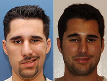
Acoustic neuroma facial paralysis affects patients in different ways, therefore it is very important to understand the type of facial paralysis in order to determine the best treatment option. Facial paralysis following acoustic neuroma removal can be mild to severe, and there is also a chance of developing synkinesis during recovery.
Acoustic neuroma facial paralysis affects patients in different ways. Therefore, it is important to understand the type of facial paralysis in order to determine the best treatment option. Facial paralysis following acoustic neuroma removal can be mild to severe, and there is also a chance of developing synkinesis during recovery.
Treating an acoustic neuroma requires expert support. Fortunately, Dr. Babak Azizzadeh of The Facial Paralysis Institute has the skills and experience to address acoustic neuroma facial paralysis. He works with acoustic neuroma facial paralysis patients to understand their symptoms. Dr. Azizzadeh then crafts a custom treatment plan designed to deliver long-lasting results.
Dr. Azizzadeh is a dual-certified and Harvard-trained facial plastic and reconstructive surgeon. He is globally recognized for his industry expertise and has performed many facial paralysis procedures over the years. Dr. Azizzadeh constantly explores ways to help acoustic neuroma facial paralysis patients, too. He is committed to helping his patients in any way possible, and to accomplish his goal, explores innovative treatment options. That way, Dr. Azizzadeh ensures each of his acoustic neuroma facial paralysis patients can accomplish their treatment goals.
What Acoustic Neuroma Surgery Options Does Dr. Azizzadeh Offer?
Dr. Azizzadeh conducts a patient evaluation to determine if an individual is a good candidate for an acoustic neuroma surgery. He performs this evaluation at his Beverly Hills office. Dr. Azizzadeh provides virtual consultations to out-of-town surgery candidates as well.
Over the years, Dr. Azizzadeh has performed many surgeries for acoustic neuromas. These treatments include:
1. Hypoglossal-Facial Nerve Transfer
A hypoglossal-facial nerve transfer involves attaching the hypoglossal (trigeminal) nerve to the gracilis muscle. The procedure helps a patient regain the ability to thrust their tongue voluntarily to move their facial muscles. It can be used to simultaneously stimulate facial muscle movement and alleviate facial numbness and paralysis.
2. Masseter to Facial Nerve Transfer
Dr. Azizzadeh may recommend a masseter to facial nerve transfer to patients who have been experiencing facial paralysis symptoms for less than three years. The procedure involves connecting the facial nerve and masseter nerve, which is used to activate the chewing muscles in the mouth. During the procedure, Dr. Azizzadeh makes an incision near the front of his patient’s ear. Dr. Azizzadeh then identifies the facial and masseter nerves and sews them together. The masseter nerve can then provide neural input into the paralyzed facial nerve to help a patient regain the ability to naturally move their facial muscles.
3. Gracilis Muscle Free Flap
A gracilis muscle free flap procedure involves taking a small portion of a patient’s inner thigh muscle and transplanting it into their face. Next, the nerve that moves the gracilis muscle (obturator nerve) is connected to a new nerve supply in the face. This allows the obturator nerve to power the gracilis muscle. As a result, a patient will be able to smile, frown, and make other facial expressions once again.
4. Temporalis Tendon Transfer
With a temporalis tendon transfer, Dr. Azizzadeh makes an incision into a patient’s temporalis muscle, which is located between the lip and cheek. He then attaches the temporalis tendon and bones to muscles in the corner of his patient’s mouth. This helps the patient improve facial symmetry.
There is no one-size-fits-all treatment to address acoustic neuromas. By partnering with Dr. Azizzadeh, a patient can explore all of the treatment options at their disposal. He or she can gain insights into myriad treatment options. Then, the patient can decide how to address their acoustic neuroma symptoms and prevent them from recurring.
Schedule an Acoustic Neuroma Treatment Consultation with Dr. Azizzadeh
At the first sign of acoustic neuroma symptoms, meet with a doctor. This ensures an individual can find out if he or she is dealing with an acoustic neuroma. Furthermore, if a neuroma is present, this individual can mitigate the problem before it causes permanent facial paralysis or other long-term health issues.
Dr. Azizzadeh dedicates the necessary time and resources to evaluate acoustic neuroma facial paralysis patients. He then works with his patients to determine the best course of action to treat their acoustic neuroma facial paralysis symptoms. Dr. Azizzadeh even responds to a patient’s acoustic neuroma facial paralysis concerns or questions to ensure this individual knows what to expect throughout treatment.
Dr. Azizzadeh is happy to meet with an individual and provide a personalized acoustic neuroma treatment recommendation. To schedule a consultation with Dr. Azizzadeh, please contact us today at (310) 657-2203.
Request your consultation with Dr. Azizzadeh today
Call us at (310) 657-2203 to schedule an appointment.
Schedule a Consultation